speakers
Alex Pollen, University of California, San Franscisco
Azahara Oliva Gonzalez, Cornell University
Barbara Treutlein, ETH Zürich
Bing Brunton, University of Washington
Bing Su, Kunming Institute of Zoology
Christopher Lowe, Stanford University
Claude Desplan, New York University
Daniel Kronauer, Rockefeller University
David Keays, University of Cambridge
Detlev Arendt, EMBL Heidelberg
Erich Jarvis, Rockefeller University
Evan Eichler, University of Washington
Franck Polleux, Columbia University
Genevieve Konopka, University of California, Los Angeles
Gilles Laurent, Max Planck Institute for Brain Research
Henrik Kaessmann, Heidelberg University (ZMBH)
Jacob Musser, Yale University
Jennifer Li, Max Planck Institute for Biological Cybernetics
Kathleen Millen, Seattle Children’s Hospital
Lauren O’Connell, Stanford
Lindy McBride, Princeton
Madeleine Lancaster, University of Cambridge
Maria Tosches, Columbia University
Nate Sawtell, Columbia University
Oliver Hobert, Columbia University
Paul Cisek, University of Montreal
Pawel Burkhardt, University of Bergen
Pierre Vanderhaegen, KU Leuven
Stein Aerts, KU Leuven/ VIB.AI
Trygve Bakken, Allen Institute
Vanessa Ruta, Rockefeller University
Postsynaptic receptors and PSD component
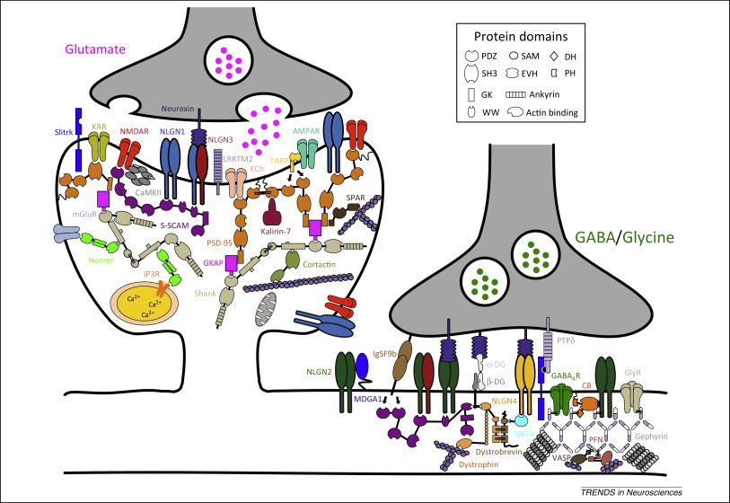
1. The Excitatory (Glutamate) Synapse
Excitatory synapses are characterized by a thick, dense network of proteins called the Postsynaptic Density (PSD). The logical flow here moves from the physical connection between neurons down to the actin skeleton that gives the spine its shape.
A. Trans-Synaptic Adhesion (The Synapse Bridge)
-
Neurexin (NRXN1-3): Presynaptic adhesion molecules that trigger postsynaptic differentiation.
-
NLGN1 & NLGN3 (NLGN1, NLGN3): Postsynaptic cell-adhesion proteins specific to excitatory synapses that align the presynaptic active zone with postsynaptic receptors.
-
Binds extracellularly to presynaptic Neurexins and intracellularly to the master scaffold PSD-95 via a PDZ-domain binding motif.
-
-
LRRTM2 (LRRTM2, Leucine-Rich Repeat Transmembrane Protein 2): A potent postsynaptic organizer that instructs the development and maintenance of excitatory synapses.
-
Binds extracellularly to presynaptic Neurexins and intracellularly to PSD-95.
-
-
Slitrk (SLITRK1-6, SLIT and NTRK-like Family Member): Postsynaptic adhesion molecules that control excitatory synapse formation and neurite outgrowth.
-
Binds trans-synaptically to presynaptic protein tyrosine phosphatases (like PTP$\sigma$).
-
B. Membrane Receptors & Channels (The Signal Receivers)
-
AMPAR (GRIA1-4, Glutamate Ionotropic Receptor AMPA Type Subunit 1-4): The primary fast-acting excitatory receptor. It opens to allow sodium influx, causing immediate depolarization.
-
Directly binds to TARPs, which ferry it to the membrane and anchor it to PSD-95.
-
-
NMDAR (GRIN1, GRIN2A-D, Glutamate Ionotropic Receptor NMDA Type Subunit 1/2): A slower, voltage-dependent receptor that allows calcium influx, critical for synaptic plasticity (learning and memory).
-
Binds directly to the PDZ domains of PSD-95 and S-SCAM (it seems only bind to NMDAR comparing to PSD-95).
-
-
KAR (GRIK1-5, Glutamate Ionotropic Receptor Kainate Type Subunit 1-5): Modulates network excitability and presynaptic neurotransmitter release.
-
mGluR (GRM1-8, Glutamate Metabotropic Receptor 1-8): G-protein coupled receptors that trigger slow, intracellular biochemical signaling cascades rather than passing ions directly.
-
Intracellularly binds directly to Homer scaffolding proteins.
-
-
KCh (Potassium Channels, various genes): Regulates the resting membrane potential and shapes the electrical action potential.
-
Binds to PSD-95 for localization at the synapse.
-
-
IP3R (ITPR1-3, Inositol 1,4,5-Trisphosphate Receptor Type 1-3): An intracellular calcium channel located on the Endoplasmic Reticulum (ER). It releases stored calcium into the spine when triggered by mGluR signaling.
-
Physically linked to surface mGluRs via the Homer scaffold.
-
C. The Core Postsynaptic Density (The Structural Scaffolds)
-
PSD-95 (DLG4, Disks Large MAGUK Scaffold Protein 4): The master organizer of the excitatory synapse. It clusters receptors and adhesion molecules at the surface.
-
Binds NMDARs, KCh, NLGN1, NLGN3, LRRTM2, TARPs, GKAP, Kalirin-7, and SPAR.
-
-
TARP (CACNG2-8, Transmembrane AMPA Receptor Regulatory Protein): Acts as an essential auxiliary subunit for AMPARs, regulating their trafficking and channel properties.
-
Links AMPA receptors directly to PSD-95.
-
-
GKAP (DLGAP1-4, DLG Associated Protein 1-4): An adaptor protein that acts as the intermediate vertical bridge in the PSD.
-
Binds the Guanylate Kinase (GK) domain of PSD-95 and connects it to Shank.
-
-
Shank (SHANK1-3, SH3 and Multiple Ankyrin Repeat Domains 1-3): A massive, multi-domain scaffolding protein that forms a deep sheet cross-linking the PSD to the actin cytoskeleton.
-
Binds GKAP, Homer, and Cortactin.
-
-
Homer (HOMER1-3, Homer Scaffold Protein 1-3): Cross-links scaffolding networks and physically connects cell-surface receptors to intracellular calcium stores.
-
Binds Shank, mGluRs, and IP3Rs.
-
-
S-SCAM (MAGI2, Membrane Associated Guanylate Kinase Inverted 2): An auxiliary scaffold that helps assemble and stabilize the PSD during early development.
-
Binds NMDARs and Neuroligins.
-
D. Cytoskeletal & Signaling Regulators (The Shape Shifters)
-
CaMKII (CAMK2A-D, Calcium/Calmodulin Dependent Protein Kinase II): A critical kinase activated by calcium influx. It alters receptor function and spine structure to strengthen the synapse (Long-Term Potentiation).
-
Binds directly to NMDARs and acts on AMPARs.
-
-
Kalirin-7 (KALRN, Kalirin RhoGEF Kinase): A signaling exchange factor that regulates the remodeling of the actin cytoskeleton, changing the physical shape of the dendritic spine.
-
Binds to PSD-95.
-
-
Cortactin (CTTN, Cortactin): Promotes the branching of actin networks, which is necessary for the dendritic spine to expand or change shape.
-
Binds to Shank and the F-actin cytoskeleton.
-
-
SPAR (SIPA1L1, Signal-Induced Proliferation-Associated 1 Like 1): A regulatory protein that reorganizes the actin cytoskeleton to control spine head size.
-
Binds to PSD-95.
-
2. The Inhibitory (GABA/Glycine) Synapse
Inhibitory synapses act to dampen electrical activity. Structurally, they are flatter and utilize a completely different set of adhesion and scaffolding molecules centered around the protein Gephyrin and the Dystrophin complex.
A. Trans-Synaptic Adhesion & Regulation
-
NLGN2 & NLGN4 (NLGN2/4, Neuroligin 2/4): Postsynaptic adhesion molecules specifically dedicated to organizing inhibitory synapses.
-
Verified Interactions: Binds presynaptic Neurexins and postsynaptic Gephyrin.
-
-
PTPdelta (PTPRD, Protein Tyrosine Phosphatase Receptor Type D): A presynaptic molecule that organizes the active zone for GABA release.
-
Verified Interactions: Binds postsynaptic partners like Slitrks or IL1RAPL1 to mediate synapse formation.
-
-
IgSF9b (IGSF9B, Immunoglobulin Superfamily Member 9B): An adhesion molecule that heavily promotes the development of inhibitory interneuron synapses.
-
Verified Interactions: Exhibits homophilic binding (binds to other IgSF9b molecules across the cleft).
-
-
MDGA1 (MDGA1, MAM Domain Containing Glycosylphosphatidylinositol Anchor 1): A unique negative regulator. It prevents over-formation of inhibitory synapses.
-
Verified Interactions: Binds in cis (on the same cell membrane) to Neuroligin-2, physically blocking it from reaching across the cleft to bind presynaptic Neurexins.
-
B. Membrane Receptors
-
GABA$_A$R (GABRA1-6, GABRB1-3, GABRG1-3, Gamma-Aminobutyric Acid Type A Receptor Subunits): The primary fast inhibitory receptor. It opens a chloride channel, hyperpolarizing the neuron and silencing it.
-
Verified Interactions: Binds directly to Gephyrin and Collybistin.
-
-
GlyR (GLRA1-4, GLRB, Glycine Receptor Subunits): The primary inhibitory receptor in the spinal cord and brainstem, also passing chloride ions.
-
Verified Interactions: Binds strongly to Gephyrin.
-
C. The Core Scaffold
-
Gephyrin (GPHN, Gephyrin, /ˈɡɛfɪrɪn/): The master scaffolding protein of the inhibitory synapse (the equivalent of PSD-95). It forms a hexagonal lattice that traps receptors at the synapse.
-
Binds GABA$_A$R, GlyR, Neuroligin-2, Collybistin, Profilin (PFN), and Mena/VASP.
-
-
Collybistin (CB) (ARHGEF9, Cdc42 Guanine Nucleotide Exchange Factor 9): A specific, essential adaptor that brings Gephyrin to the cell membrane to form clusters.
-
Binds Gephyrin and specific GABA$_A$ receptor subtypes.
-
D. The Dystrophin-Glycoprotein Complex & Actin Linkers (Structural Anchors)
-
alpha-DG & beta-DG (DAG1, Dystroglycan 1. They call is Dystro- prefix since it is most important in musle dystrophy): A two-part receptor complex that physically connects the outside environment to the inside skeleton, providing immense structural stability to the synapse.
-
alpha-DG binds extracellular matrix proteins (like Neurexin); beta-DG binds intracellular Dystrophin.
-
-
Dystrophin (DMD, Dystrophin): A massive rod-like protein that anchors the membrane complexes deep into the neuron's structural framework.
-
Binds beta-Dystroglycan, Actin filaments, Dystrobrevin, and Syntrophin.
-
-
Dystrobrevin (DTNA/B, Dystrobrevin Alpha/Beta): Works alongside Dystrophin to stabilize the DGC complex.
-
Binds Dystrophin and Syntrophin.
-
-
SNTA (Syntrophin $\alpha$) (SNTA1, Syntrophin Alpha 1): An adaptor protein that links the structural Dystrophin complex to localized signaling molecules.
-
Binds Dystrophin and Dystrobrevin.
-
-
VASP (VASP, Vasodilator-Stimulated Phosphoprotein): A protein that regulates the length and dynamics of actin filaments.
-
Binds Gephyrin and Profilin.
-
-
PFN (Profilin) (PFN1-4, Profilin 1-4): Binds to individual actin building blocks (monomers) and shuttles them to areas where the cytoskeleton needs to grow.
-
Binds Gephyrin, VASP, and Actin monomers.
-
Neurexin and Neuroligin funcion
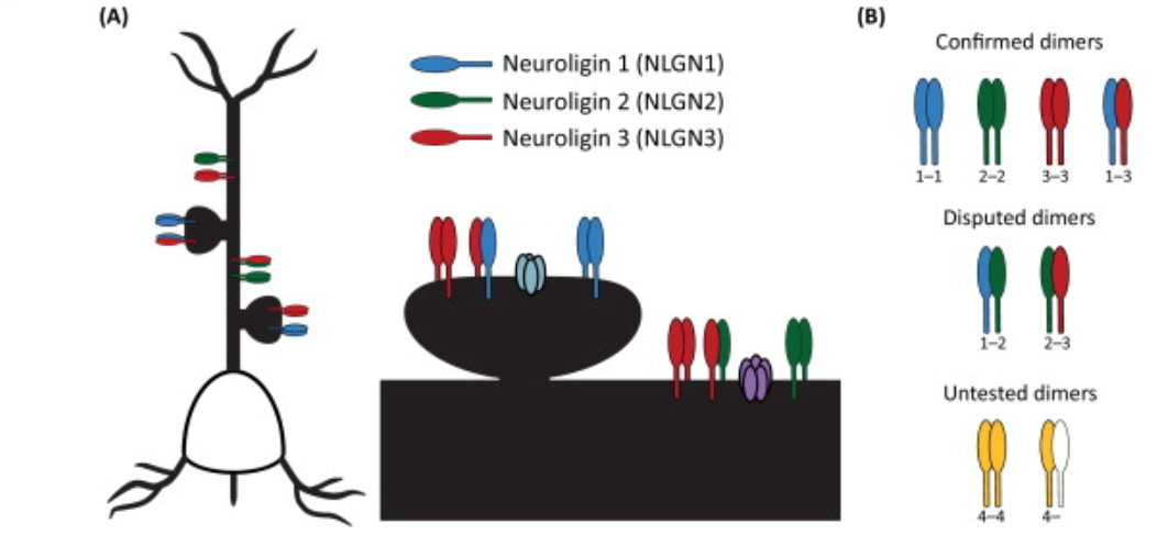
NLGN1 is localized to excitatory (glutamatergic) synapses; NLGN2 is localized to inhibitory (GABAergic) synapses; and NLGN3 is localized to both excitatory and inhibitory synapses. NLGNs tend to form dimers, either homo or hetro (fixed pattern, NLGN1 + NLGN3 for excitatory synapse or NLGN2 + NLGN3 for inhibitory synapse).
Severual important functions
1, Presynaptic neurexin and postsynapic neuroligin are glue to stablize and sustain the sypase cleft. It is their structural function.
2, The intracellular tails of neuroligins bind to specific scaffolding molecules to build the postsynaptic density. For example, NLGN1 bind to PSD-95 at excitatory (glutamatergic) synapses , and NLGN2 recruits gephyrin and collybistin to organize inhibitory (GABAergic) postsynaptic specializations.
3, Extracellular domain of neuroligin 1 (NLGN1) can interact directly with NMDA-type glutamate receptors. In this role, NLGN1 acts as a shuttle to deliver these receptors to the synapse, directly affecting synaptic strength rather than relying solely on intracellular scaffolds.
4, Its also initiate and promote synapse formation (synaptogenesis). Many overexpression and KO experiment have shown that increasing neuroligin expression results in more synapses, whereas reducing neuroligin expression results in fewer synapses. This physical adhesion is one of the earliest event in synaptogenesis. (only the binding of NRXN and NLGN, without any neurotransmitter being release or received. Actually they recruit the SNARE and PSD to make the release and receive happen)
5, Synapses can still form following the triple knockout of NLGNs1–3 (showing redundency of adhesion pair during synaptogenesis). But interesting, tissue level knock out always show a very mild phenotype, where OE shows dramatic effet. A famous experiment (Nat. Neuro 2012) shows only a mixture of NLGN1-/- and wild type cells shows a synaptic effet. suggesting a cell-to-cell competitive plasticity.
6, Synaptic activity triggers calcium influx, prompting the kinase CaMKII to transiently phosphorylate NLGN1 (at residue T739). This phosphorylation dramatically increases the surface expression of NLGN1, promoting the creation of new synapses in response to neural activity.
7, Combine the point 5 and 6, these mechanism allows highly active cells to gain more synapses at the expense of less active neighbors (a initial difference in NLGNs density -> differ in response the calcium influx-related plasitcity -> more NLGN being recruited in active cells -> initial difference is amplified), helping non-uniform neural networks develop specialized, over-connected "hubs" of activity. (active cells have more synapse).
8, NLGN1 is inhibited by MDGA2, where NLGN2 is inhibited by MDGA1 (MDGAs bind NLGNs in the way that as neurexin do to block its function)
Some further details
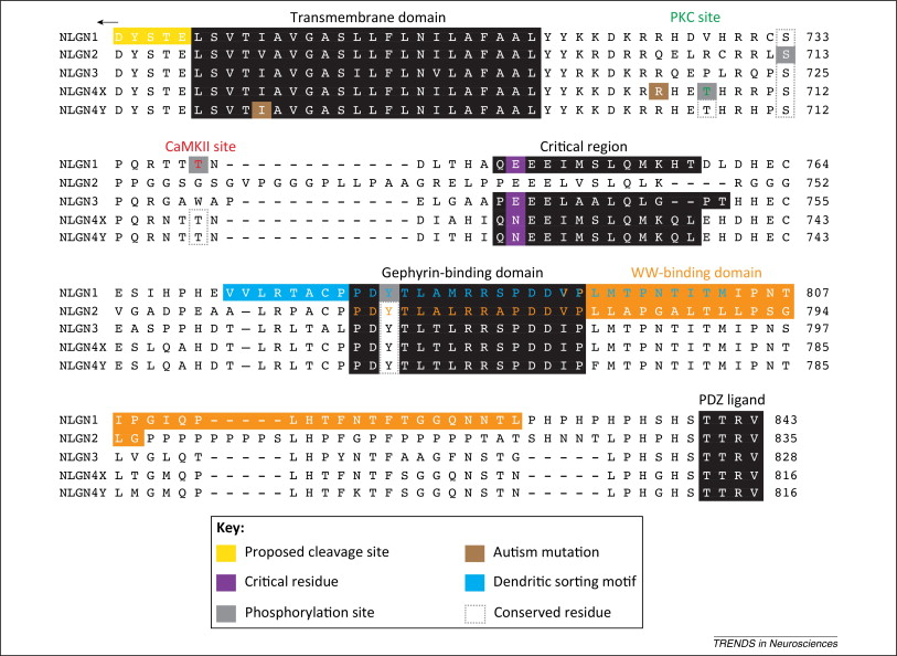
NLGNs has PDZ ligand c-terminal (to bind PSD95 or s-SCAM in excitatory postsynapse), also they have binding region to bind gephyrin in inhibitory postsynapse.
Complete walkthrough of synaptic related term
A complete workthrough of synaptic biology.
Before neurotransmitter release can occur, the presynaptic bouton must undergo depolarization, followed by a massive, localized influx of calcium (Ca2+) that triggers exocytosis.
1. Action Potential Propagation & Ion Channel Regulation
The action potential is driven by Voltage-Gated Sodium Channels (VGSCs) and repolarized by Potassium channels.
Na+ sodium ion or sodium cation (/ˈkætˌaɪ.ɑːn/, combines "cat-" as in cat and "-ion" as in eye-on. The negative ion is called anion, /ˈæn.aɪ.ən/, consists of three syllables: "an-" (as in pan), "-i-" (as in eye), and "-on" (as in upon), with the primary stress on the first syllable)
Cl- chloride (/ˈklɔː.raɪd/) ion
Mg2+ magnesium ( /mæɡˈniːziəm/) ion
-
FGF12 & FGF14: These are crucial auxiliary proteins that bind to the intracellular domains of VGSCs (like *SCN1A/SCN2A*, Nav1.1 and Nav1.2), regulating their gating properties to ensure the fidelity of high-frequency firing. FGF11–FGF14 are different from other FGFs, which are called intracellular fibroblast growth factor (iFGF) family and basically acting as regulators of voltage-gated sodium and calcium channels in the brain, rather than as secreted growth factors.
-
KCNA (drosophila homolog shaker, sh, the first potassium votage gated gene, found by Yuh Nung Jan), KCNIP4, DPP10: Auxiliary subunits for A-type voltage-gated potassium channels (Kv4.x). They dictate the rapid repolarization of the membrane, preventing excitotoxicity and preparing the terminal for the next spike.
2. Excitation-Secretion Coupling in presynapse (The SNARE Machinery)
This is the core mechanism of synaptic vesicle release. Vesicles do not simply burst; they are mechanically forced to fuse with the membrane by a complex of proteins.
- *CACNA1A/B (Voltage-Gated Calcium Channels): Depolarization opens these channels, creating Ca2+ microdomains at the active zone.
- The SNARE Core (*STX1A, *SNAP25, *VAMP2): These three proteins zipper together with strong force, pulling the vesicle membrane and the presynaptic membrane together (the structural component to tie vesicle to the ready zone). Their fusion results in quantal release of neurotransmitters (like glutamate or GABA. btw GAD1 is a enzyme which transfer glutamate to GABA, which is crazy).
- *SYT1 (Synaptotagmin-1): The fundamental calcium sensor on the vesicle. When Ca2+ binds to SYT1, it triggers the final fusion event.
-
SYN2 (Synapsin II): Tethers the "reserve pool" of vesicles to the actin cytoskeleton. Upon depolarization, Ca2+ activates kinases that phosphorylate Synapsin, detaching vesicles so they can join the "readily releasable pool" at the active zone. While SYN1/2 are primarily responsible for trafficking and organizing the reserve pool of vesicles, SYT1 acts as the calcium sensor that triggers the immediate fusion of these vesicles. All of them are the regulator, and not actually invovled in the SNARE complex (the structural compoenent of the tie)
-
CNTNAP4: Specialized in regulating the exocytosis kinetics specifically for GABAergic and dopaminergic vesicles.
-
NSG2 & LRRK2: Govern the endosomal sorting and recycling of these vesicles post-fusion, ensuring the terminal maintains its vesicular supply.
Phase 2: Trans-Synaptic Alignment & Cleft Clearance
The pre- and post-synaptic densities are not just floating near each other; they are physically anchored to form highly precise "nanocolumns." This ensures the site of vesicle fusion perfectly aligns with the highest concentration of post-synaptic receptors.
-
The Synaptic Adhesion Matrix: NRXN1 (Presynaptic Neurexin) spans the cleft to bind NLGN1/2 (Postsynaptic Neuroligin). This interaction recruits the vesicles on one side and the receptors on the other. LRRTM4 acts synergistically with Neuroligins to stabilize excitatory synapses.
-
MDGA2: Functions as a critical steric inhibitor. By binding to Neuroligins (since it has a structure mimiking neurexin), it blocks the Neurexin-Neuroligin interaction, providing a regulatory "brake" on synapse formation to prevent hyper-connectivity.
-
Axon Guidance & Cleft Matrix: SLIT1 and SPOCK1 maintain the structural integrity of the extracellular space and guide synaptic target recognition.
-
Cleft Clearance (*SLC1A2 / EAAT2): After release, neurotransmitters must be rapidly cleared to prevent receptor desensitization. Glial cells and neurons use specific transporters to pump glutamate and GABA out of the cleft.
Phase 3: Post-Synaptic Reception (Ionotropic vs. Metabotropic)
This is where the chemical signal is transduced back into cellular logic. Receptors are divided into two fundamental classes based on their mechanism of action.
A. Ionotropic Receptors (Ligand-Gated Ion Channels)
Ionotropic /aɪˌɑːnəˈtrɑːpɪk/
These mediate fast synaptic transmission (milliseconds). When a neurotransmitter binds, the protein physically changes shape, opening a pore that allows ions to flow across the membrane, creating an Excitatory or Inhibitory Post-Synaptic Potential (EPSP or IPSP).
-
AMPA Receptors (glutamate ionotropic receptor AMPA type subunit 1-4, "GRIA" prefix, GRIA1-4): The workhorses of fast excitatory transmission. Glutamate binding opens the pore to Na+, instantly depolarizing the spine.
-
NMDA Receptors (glutamate ionotropic receptor NMDA, "GIRN" prefix, GRIN2B, GRIN3A, GRIN1): The "coincidence detectors" essential for synaptic plasticity. At resting membrane potential, their pore is blocked by a Magnesium (Mg2+) ion. They only open if two conditions are met: glutamate binds, and the membrane is already depolarized (usually by AMPA receptors) to repel the Mg2+ block. When open, they allow heavy influx of Ca2+, which acts as a powerful intracellular signal. NMDA receptor contains subunit as GluN1s, GluN2s and GluN3s (most commonly it at least include GluN1 and GluN2). The choice to include which subunits into the NMDA receptor determine receptor kinetics, magnesium sensitivity.
GluN2B is prominent in the early postnatal brain, mediating longer-lasting synaptic currents. GluN2A ratio increases in number during development, replacing GluN2B, and is associated with faster kinetics. The shift from GluN2B to GluN2A is crucial for synaptic maturation. -
Kainate Receptors (/'kaɪneɪt/, GRIK2): Another class of fast glutamate receptors, but unique because they often exist presynaptically as well as postsynaptically, where they act to auto-regulate neurotransmitter release.
-
GABA-A Receptors (GABRB2, *GABRA1, *GABRG2): The primary mediators of fast inhibition. GABA binding opens a Chloride (Cl-) pore. The influx of negative Cl- ions hyperpolarizes the cell, shunting incoming excitatory signals.
B. Metabotropic Receptors (G-Protein Coupled Receptors - GPCRs)
These mediate slow, neuromodulatory transmission (seconds to minutes). They do not have an ion pore. Instead, neurotransmitter binding activates an intracellular G-protein, triggering secondary messenger cascades (like cAMP or IP3/DAG) that fundamentally alter the neuron's biochemical state.
-
Metabotropic Glutamate Receptors (mGluRs):
-
Group I (*GRM1 glutamate metabotropic receptor, *GRM5): Postsynaptic. Coupled to Gq proteins, they trigger intracellular Ca2+ release from the endoplasmic reticulum, modulating excitability.
-
Group II/III (GRM7, *GRM2-4, 6, 8): Primarily presynaptic auto-receptors. Coupled to Gi/o proteins. When glutamate spills over into the perisynaptic space, it binds GRM7, which suppresses adenylyl cyclase, inhibits presynaptic Ca2+ channels, and scales back further glutamate release (a negative feedback loop).
-
-
Metabotropic GABA Receptors (*GABBR1, *GABBR2): Presynaptic and postsynaptic slow inhibitors. They act via Gi/o proteins to open specific potassium channels (GIRKs) and inhibit presynaptic calcium channels, creating a long-lasting inhibitory tone.
-
GPR158: An orphan GPCR that acts as a structural organizer for intracellular signaling pathways, heavily involved in stress responses and mood regulation at the postsynaptic density.
Phase 4: The Post-Synaptic Density (PSD) Scaffolding
Receptors are anchored directly beneath the presynaptic release sites by a dense, gel-like matrix of scaffolding proteins.
-
DLG4 (PSD-95): The master architect of the excitatory PSD. It contains PDZ domains that physically hold NMDA and AMPA receptors in the membrane and link them to intracellular signaling proteins. DLG2 (top hit in my study), along with DLG3 (SAP-102), are scaffolding molecules that localize at the postsynaptic density (PSD) of excitatory synapses. While DLG4-related synaptopathy is well-established as an ultra-rare genetic disorder, DLG2 deficiencies have been linked to a very broad NDDs including autism-related behavioral changes.
-
LRRC7 (Densin-180): A transmembrane protein localized to the PSD that binds tightly to PSD-95 and CaMKII, helping cluster the receptor apparatus.
-
*SHANK & *HOMER: Deeper scaffolding proteins that link PSD-95 to the actin cytoskeleton and to intracellular calcium stores. Shank proteins directly bind to both Homer and the PSD-95 complex (via GKAP/SAPAP), forming a "high-order polymerized complex" or "mesh-like network" that cross-links mGluRs and NMDARs.
-
GPHN (Gephyrin): The inhibitory equivalent to PSD-95. It anchors GABA-A (GABRB2) receptors at inhibitory synapses.
Phase 5: Synaptic Plasticity, Homeostasis, & Metabolism
The synapse is a living computational unit. High-frequency stimulation leads to Long-Term Potentiation (LTP)—a physical strengthening of the synapse—which is the molecular basis of learning and memory.
1. Intracellular Signaling & Plasticity
-
CAMK2A (CaMKII): When NMDA receptors let Ca2+ in, CaMKII is activated. It is a critical kinase that phosphorylates AMPA receptors (making them conduct more Na+) and drives the insertion of new AMPA receptors into the membrane, strengthening the synapse.
-
Actin Remodeling: To accommodate more receptors, the dendritic spine must physically grow. COBL and TRIM67 regulate actin filament assembly, while NAV2 and GPRIN3 guide neurite and spine outgrowth.
-
Extracellular Remodeling: PLAT (Tissue Plasminogen Activator, highly express in human OPC): Released during intense synaptic activity, it cleaves extracellular matrix proteins, physically making room for spines to enlarge during late-phase LTP.
2. Ion Homeostasis & Energy Support The intense ionic flux of synaptic transmission demands rigorous cleanup and massive energy expenditure.
-
SLC8A1 & SLC8A3 (NCX): Sodium-Calcium exchangers. Following NMDA receptor activation, these transport proteins rapidly pump Ca2+ out of the spine (exchanging it for Na+) to restore baseline resting levels and prevent excitotoxic cell death.
-
Metabolism & Lipids: The continuous fusion and recycling of vesicles requires immense lipid turnover. MFSD2A transports crucial omega-3 fatty acids (DHA) across the blood-brain barrier for membrane synthesis. PLA2G7 (phospholipase), LPL (lipoprotein lipase), and RBP4 handle lipid metabolism. SLC2A13 transports myo-inositol, a precursor for the PIP2 lipid signaling pathway critical for vesicle exocytosis.
3. Transcriptomic Control (The Long Game) For long-term changes, the synapse must request new proteins from the nucleus.
-
Alternative Splicing: RBFOX1, KHDRBS2, MATR3, and RALYL are RNA-binding proteins. Neurons use alternative splicing to create highly specific isoforms of receptors and channels. For instance, RBFOX1 regulates the splicing of NMDA receptors and voltage-gated channels, fundamentally dictating the long-term electrophysiological properties of the neuron based on its history of activity.
Negative binomial and GLM in SCT normalization
1, Why GLM beats the Simple Ratio (Global Scaling even in mean value)
The traditional standard RNA assay (NormalizeData) uses a "Simple Ratio" approach: it divides a gene's count by the total UMIs in that cell, multiplies by a constant (like 10,000), and takes the log.
This relies on a massive, flawed assumption: strict linear proportionality.
It assumes that if Cell A is sequenced twice as deeply as Cell B, every single gene in Cell A will have exactly twice the counts of Cell B. In the messy reality of single-cell biology, this isn't true for two main reasons:
The "Squeezing" Effect on Abundant Genes: A few highly expressed genes (like ribosomal or mitochondrial genes) often eat up a massive fraction of your sequencing reads. If a cell happens to capture a lot of these, the total UMI goes way up.
Non-linear Dropouts: Low-abundance genes don't scale linearly with sequencing depth; they are heavily affected by technical dropouts (zeros).
On the other hands, GLM treat each gene seperately, fitting a curve in a scatter plot (x is UMI depth, y is expected expression), and take the medium UMI across cells to be a "standard cell", recalculate each gene expression when they are sequenced with the medium depth.
2, Why GLM with negative binomial beats GLM with simple OLS (ordinary linear regression, global scaling in variance)
It is basically because, in the above scatter plot, a high UMI depth cells is typically very nosiy and have a very very outlier signal. However, OLS use least sequre to penalize curve fitting, means a point with low x value and point with high x value, the least square are treated equally (for some reason this means the error probability follows normal distribution). This is very unhealty.
On the other hand, GLM with negative binomial does not calculate lest square. It first try thoudands of possible curve, and for each, mesure the error (distance between observed value with expected value). However, the tolerance of error is different based on values x coordinate (a low x value observation means strict error tolerance, a high x value observation means loose tolerance). It calculate probablity for each point (tolerance is dependent with total umi depth), and multiple all probabilty (from each point) to get a overall probability for the test curve. The curve with the highest probability wins.
Actually negative binomial is a modified version of Poisson (/pwəˈsoʊn/. It is not pronounced like the English word "poison.") (to be allowed to have a higher variance tolerance than poisson distribution).
The golden rule of the Poisson distribution is that the Mean equals the Variance ($\mu = \sigma^2$).

The variance formula for a Negative Binomial distribution is:

You can see, the Var is further added with theta controled part. It can be lot larger than the normal poisson distribution. Poisson distribution is also a tolerance dependent since its variance scale with mean but less powerful than NB
Here is a toy numeric walkthrough
Imagine we sequence 3 cells and look at Gene X. We will simplify sequencing depth to a basic multiplier (1x, 2x, 3x depth).
-
Cell 1: Depth = 1. Gene X count = 2.
-
Cell 2: Depth = 2. Gene X count = 4.
-
Cell 3: Depth = 3. Gene X count = 20. (This is our biological "burst" or overdispersed outlier).
The True Biological Baseline: Looking at Cell 1 and Cell 2, the true baseline rate of this gene is exactly 2 counts per 1 unit of depth. (Expected Count = 2 $\times$ Depth). Cell 3 just had a random burst of transcription.
Let's test two candidate lines to see which one the algorithms choose as the "best fit":
-
Line A (The True Baseline): Expected Count = $2 \times$ Depth
-
Line B (The Skewed Line): Expected Count = $5 \times$ Depth (A steeper line trying to get closer to the outlier).
Method 1: Ordinary Least Squares (OLS)
OLS assumes errors follow a Normal distribution. It evaluates a line by calculating the Sum of Squared Errors (SSE). It wants the lowest score possible.
Testing Line A (Y = 2X)
-
Cell 1 Error: Expected 2, got 2. $(2 - 2)^2 = 0$
-
Cell 2 Error: Expected 4, got 4. $(4 - 4)^2 = 0$
-
Cell 3 Error: Expected 6, got 20. $(20 - 6)^2 = 196$
-
Total OLS Score for Line A = 196
Testing Line B (Y = 5X)
-
Cell 1 Error: Expected 5, got 2. $(2 - 5)^2 = 9$
-
Cell 2 Error: Expected 10, got 4. $(4 - 10)^2 = 36$
-
Cell 3 Error: Expected 15, got 20. $(20 - 15)^2 = 25$
-
Total OLS Score for Line B = 70
The OLS Conclusion: Because 70 is much lower than 196, OLS chooses Line B as the best fit. Why it failed: Squaring the error of the bursty cell $(14^2 = 196)$ created such a massive penalty that the algorithm was forced to abandon the true baseline of Cells 1 and 2 just to make that single penalty smaller.
Method 2: Maximum Likelihood Estimation (with NB)
MLE evaluates a line by calculating Probability. It asks the Negative Binomial distribution: "If this line is the true mean, what is the probability of actually seeing the counts we observed?" It multiplies these probabilities together and wants the highest score possible.
Crucial NB Rule: In a Negative Binomial distribution, as the expected mean gets larger, the expected variance (spread) gets exponentially larger.
Testing Line A (Y = 2X)
-
Cell 1 (Expected 2, got 2): Variance is low here. Hitting the expectation exactly gives a high probability. Let's say $P = 0.30$.
-
Cell 2 (Expected 4, got 4): Again, hitting it exactly. $P = 0.20$.
-
Cell 3 (Expected 6, got 20): Under a Normal distribution, being off by 14 is impossible. But the NB distribution knows this is single-cell data! It says, "At an expected mean of 6, variance is starting to blow up. A burst of 20 is rare, but entirely possible." Let's say $P = 0.04$.
-
Total MLE Score for Line A: 0.30 x 0.20 x 0.04 = 0.0024
Testing Line B (Y = 5X)
-
Cell 1 (Expected 5, got 2): Variance is supposed to be tight at low depth. Missing by 3 is heavily penalized. $P = 0.05$.
-
Cell 2 (Expected 10, got 4): Missing by 6 is heavily penalized. $P = 0.01$.
-
Cell 3 (Expected 15, got 20): Close to the expected mean, so probability is decent. $P = 0.10$.
-
Total MLE Score for Line B: 0.05 x 0.01 x 0.10 = 0.00005
The NB MLE Conclusion:
Because 0.0024 is much higher than 0.00005, the GLM chooses Line A as the best fit.
Microcephaly, MCPH and centriolar / ciliary function
Microcephaly is a rare brain disorder defined by reduced brain size while maintaining an overall normal architecture, with an occipitofrontal head circumference smaller than at least 2 or 3 standard deviations below the mean for age, gender, and ethnicity.
Microcephaly can be caused by a number of genetic and environmental factors, including, but not limited to, in utero virus infections (e.g., Zika viru2), teratogen exposure (e.g., fetal alcohol syndrome) and metabolic conditions (e.g., maternal phenylketonuria and Amish type microcephaly5,6). Genetically conditioned primary microcephaly is termed microcephaly primary hereditary /həˈrɛdɪˌtɛri/ (MCPH), which is typically inherited in an autosomal recessive manner and primarily affects the development of the neocortex.
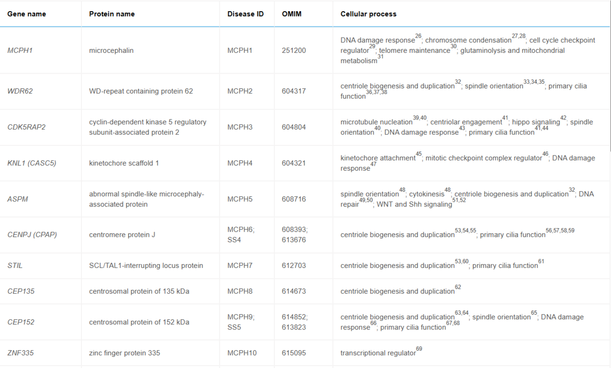
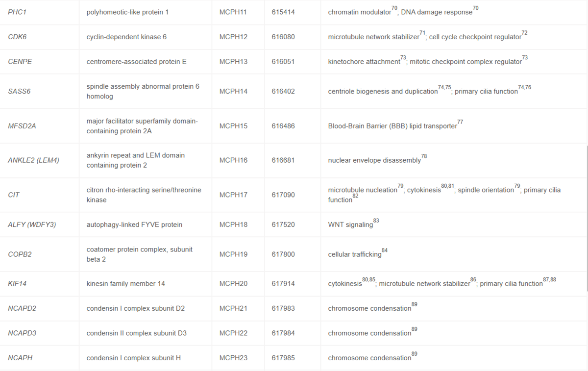
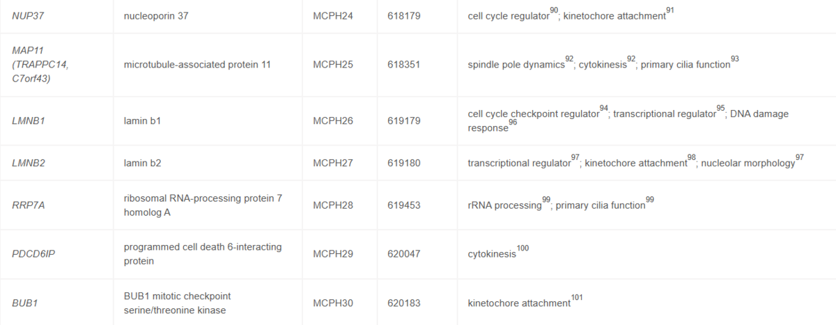
To date, at least 30 genes are implicated in MCPH, according to the OMIM database: MCPH1, WDR62, CDK5RAP2, KNL1, ASPM, CENPJ, STIL, CEP135, CEP152, ZNF335, PHC1, CDK6, CENPE, SASS6, MFSD2A, ANKLE2, CIT, WDFY3, COPB2, KIF14, NCAPD2, NCAPD3, NCAPH, NUP37, MAP11, LMNB1, LMNB2, RRP7A, PDCD6IP, and BUB1 (MCPH1-30, respectively).
These genes are basically invovled in either centriolar or ciliary function. MCPH isself is widely recognized as a "ciliopathy /ˌsɪliˈɑːpəθi/-spectrum" disorder or a disease of the centrosome, as the majority of MCPH-associated genes encode proteins that localize to the centrosome/centrioles, which are essential for centriole duplication, spindle organization, and the formation of primary cilia.
Variants in the Assembly factor for spindle microtubules (ASPM/MCPH5) gene are the most frequent cause of MCPH. ASPM regulates multiple essential cellular processes. Studies of the ASPM ortholog asp in Drosophila melanogaster revealed a role in mitotic processes, including focusing of spindle poles, centrosome attachment, and chromosome segregation. In mammalian cells, ASPM also localizes at spindle poles, where it influences spindle orientation and centrosome attachment. Additionally, an involvement of ASPM in centriole duplication at the mother centriole was demonstrated. ASPM also modulates key signaling pathways, including Wingless-related integration site (WNT) and Sonic hedgehog (Shh), which regulate cell proliferation and differentiation.
Cilia function related with signaling pathway
Ciliogenesis has three parameters. Cilium length and cilium frequency (ratio of cells bearing cilium in cell population) are positve indicators, meaning a stable, strong cilium. Cilium reabsorption speed is a negative indicators, meaning a dynamics, unstable and frequently disassemble cilium structure.
Ciliogenesis is positively correlated with SHH signaling and negatively correlated with mTOR signaling.
Ciliogenesis itself is exclusive with cell division and brake cell cycle. Longer cilium means longer G1 phase. Therefore, the mTOR pathway is straightforwad, but SHH pathway has counteracting part.
SHH brief: When SHH (Hedgehog ligand) binds to Patched-1 (PTCH1, a G protein-coupled receptor-like protein) at the base of the cilium, it allows Smoothened (SMO) to enrich within the ciliary membrane, activating the GLI transcription factors.
PTCH1 -inhibit-> SMO
SMO functions as a positive regulator of the Hedgehog pathway. It sits at the cell surface (or in the primary cilium in vertebrates) and is normally repressed by PTCH1.
A longer cilium means a larger membrane surface area and a greater volume of intraflagellar transport (IFT). This typically results in hyper-sensitivity to SHH. Even low concentrations of ambient SHH will trigger a massive accumulation of SMO, leading to an amplification of downstream targets like Gli1. In the developing cortex, this might alter the delicate balance of dorsal-ventral patterning or hyper-stimulate progenitor proliferation (counteracting the G1 delay usually caused by longer absorption time from a longer cilium, making the final phenotype highly dependent on the exact spatial location of the cell).
mTOR brief: The primary cilium actually acts as a brake on the mTORC1 pathway. The basal body of the cilium is loaded with LKB1 and AMPK. When the cilium is fully formed and sensing flow/chemical gradients, this LKB1/AMPK axis actively represses mTOR to prevent premature protein synthesis and cell growth, keeping the progenitor in a tightly controlled metabolic state.
If a progenitor maintains a long, highly stable primary cilium, you would expect reduced global mTOR activity. Conversely, if you force the rapid resorption of the cilium, the AMPK brake is released, mTOR spikes, cell size increases, and the cell rushes into mitosis.
Several famous manupulation results:
In vivo:
Ciliogenesis can be prevented by targeting key IFT (intraflagellar transport, the transport occurs inside the cilia throuogh axoneme) components such as IFT88, a core member of the IFT-B complex, or KIF3A, a kinesin-2 subunit required for IFT.
Conditional knockout (KO) of IFT88 or KIF3A from embryonic day 11 (E11) in the mouse resulted in complete loss of cilia by E14.5, accompanied by ventricular enlargement, reduced cortical thickness, apical domain expansion of aRGCs, spindle misorientation, and impaired mammalian target of rapamycin (mTOR) signaling. Remarkably, a single injection of rapamycin at E12.5 was sufficient to rescue cortical abnormalities, implicating primary cilia-dependent mTOR signaling in normal corticogenesis. Consistently, KIF3A knockdown (KD) by short hairpin RNA (shRNA) in cultured E14 mouse brain slices arrested NSCs in the VZ/SVZ at an undifferentiated state. KIF3A KD also delayed RGC cell-cycle progression and interkinetic nuclear migration after mitosis. These defects were rescued by overexpression of the Hedgehog (Hh) pathway transcription factor GLI2, which enhanced Cyclin D1 expression. Because Hh signaling is bona fide regulated by primary cilia in vertebrates, these findings support the conclusion that cilia-mediated Hh signaling promotes cell-cycle progression in aRGC. Interestingly, not all IFT88 conditional KO studies reported strong phenotypes in the E14 mouse brain,149 pointing to the importance of KO timing for ciliary function. Together, these findings establish primary cilia as regulators of aRGC expansion and corticogenesis through pathways including mTOR and Hh signaling.
In vitro:
CROCCP2 overexpression in either mouse cortex or human organoids results in a shorter cilium length, lower cilium frequency (lower raio of cells bearing cilium), higher signal of mTOR (p6S positive) and a higher ratio of proliferative TBR2 cells.
