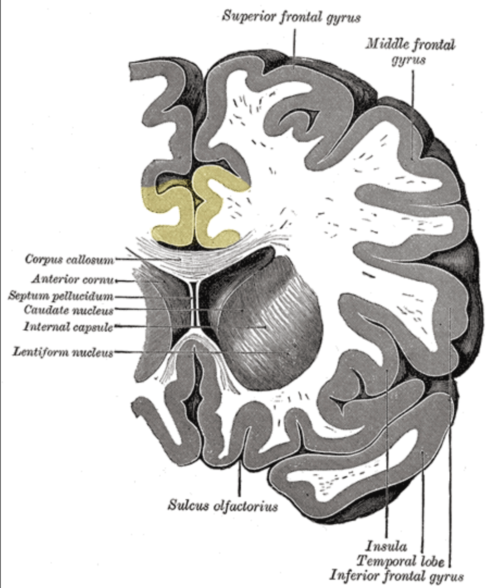
Basal ganglia in general
Most of the neurons inside it are inhibitory GABA neurons or modulatory dopaminergic neurons.
The basal ganglia (BG), or basal nuclei, are a group of subcortical nuclei, of varied origin, in the brains of vertebrates. The basal ganglia are situated at the base of the forebrain and top of the midbrain. Basal ganglia are strongly interconnected with the cerebral cortex, thalamus, and brainstem, as well as several other brain areas. The basal ganglia are associated with a variety of functions, including control of voluntary motor movements, procedural learning, habit learning, conditional learning,[1] eye movements, cognition,[2] and emotion. Popular theories implicate the basal ganglia primarily in action selection – in helping to decide which of several possible behaviors to execute at any given time. In more specific terms, the basal ganglia's primary function is likely to control and regulate activities of the motor and premotor cortical areas so that voluntary movements can be performed smoothly.
Movement disorders include, most notably Parkinson's disease, which involves degeneration of the dopamine-producing cells in the substantia nigra; Huntington's disease, which primarily involves damage to the striatum;[2][4] dystonia; and more rarely hemiballismus.
Striatum
The striatum is a subcortical structure generally divided into the dorsal striatum and ventral striatum, although a medial lateral classification has been suggested to be more relevant behaviorally and is being more widely used.
The striatum is composed mostly of medium spiny neurons (MSNs). These GABAergic neurons project to the external (lateral) globus pallidus and internal (medial) globus pallidus as well as the substantia nigra pars reticulata. The projections into the globus pallidus and substantia nigra are primarily dopaminergic, although enkephalin, dynorphin and substance P are expressed. The striatum also contains interneurons that are classified into nitrergic neurons (due to use of nitric oxide as a neurotransmitter), tonically active (i.e. constantly releasing neurotransmitter unless inhibited) cholinergic interneurons, parvalbumin-expressing neurons and calretinin-expressing neurons.[18] The dorsal striatum receives significant glutamatergic inputs from the cortex, as well as dopaminergic inputs from the substantia nigra pars compacta. The dorsal striatum is generally considered to be involved in sensorimotor activities. The ventral striatum receives glutamatergic inputs from the limbic areas as well as dopaminergic inputs from the VTA, via the mesolimbic pathway. The ventral striatum is believed to play a role in reward and other limbic functions.[19] The dorsal striatum is divided into the caudate and putamen by the internal capsule while the ventral striatum is composed of the nucleus accumbens and olfactory tubercle.[20][21] The caudate has three primary regions of connectivity, with the head of the caudate demonstrating connectivity to the prefrontal cortex, cingulate cortex (part of the cortex, superior to the corpus callosum and caudate nucleus) and amygdala (the most anterior part of caudate).

The cingulate cortex is a part of the brain situated in the medial aspect of the cerebral cortex. The cingulate cortex includes the entire cingulate gyrus, which lies immediately above the corpus callosum, and the continuation of this in the cingulate sulcus. The cingulate cortex is usually considered part of the limbic lobe.

Pallidum
The pallidum consists of a large structure called the globus pallidus ("pale globe") together with a smaller ventral extension called the ventral pallidum. The globus pallidus appears as a single neural mass, but can be divided into two functionally distinct parts, called the internal (or medial) and external (lateral) segments, abbreviated GPi and GPe. Both segments contain primarily GABAergic neurons, which therefore have inhibitory effects on their targets. The two segments participate in distinct neural circuits. The GPe receives input mainly from the striatum, and projects to the subthalamic nucleus. The GPi receives signals from the striatum via the "direct" and "indirect" pathways. Pallidal neurons operate using a disinhibition principle. These neurons fire at steady high rates in the absence of input, and signals from the striatum cause them to pause or reduce their rate of firing. Because pallidal neurons themselves have inhibitory effects on their targets, the net effect of striatal input to the pallidum is a reduction of the tonic inhibition (i.e. constantly releasing inhibitory neurotransmitter unless inhibited. Also has tonic active which has adverse meaning) exerted by pallidal cells on their targets (disinhibition) with an increased rate of firing in the targets.
Substantia nigra
The substantia nigra is a midbrain gray matter portion of the basal ganglia that has two parts – the pars compacta (means this part are highly dense and compact) (SNc) and the pars reticulata (means less dense) (SNr). SNr often works in unison with GPi, and the SNr-GPi complex inhibits the thalamus. Substantia nigra pars compacta (SNc) however, produces the neurotransmitter dopamine, which is very significant in maintaining balance in the striatal pathway. The circuit portion below explains the role and circuit connections of each of the components of the basal ganglia.

Subthalamic nucleus
The subthalamic nucleus is a diencephalic gray matter portion of the basal ganglia, and the only portion of the ganglia that produces an excitatory neurotransmitter, glutamate. The role of the subthalamic nucleus is to stimulate the SNr-GPi complex and it is part of the indirect pathway. The subthalamic nucleus receives inhibitory input from the external part of the globus pallidus and sends excitatory input to the GPi.